
Fig. 3.
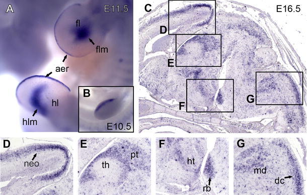
Neph2 expression as revealed by in situ hybridization analysis. Magnification of fore- and hindlimb buds of an E11.5 mouse embryo (a). Embryo is shown from the left, the back directed to the right. The insert shows earlier stage (E10.5) of fore limb development (b). Sagittal sections of mouse brain at stage E16.5 (c). Higher magnification of neocortex (d), diencephalon (e), pons (f), and oblong medulla (g) as indicated by boxes. aer apical ectodermal ridge, dc dorsal column tract, fl fore limb; flm fore limb mesenchyme, hl hind limb, hlm hind limb mesenchyme, ht hypothalamus, md medulla, neo neocortex, pt pretectum, rb rhombomere basal plate