Figure 6.
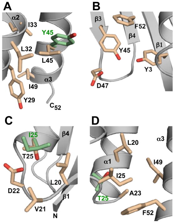
Structural basis for switching between 3α and 4β+α folds. (A) A representative NMR structure of GA98 showing L45 and nearest neighbors (pale orange) described in the text. The side chain conformation of Y45 (green) in the GB98-T25I CS-Rosetta structure is superimposed for comparison purposes. (B) GB98 NMR structure highlighting Y45 and adjacent amino acids. (C) NMR structure of GB98 showing T25 and surrounding residues (pale orange). The I25 side chain (green) in the NMR structure of GB98-T25I,L20A is superimposed for comparison. (D) CS-Rosetta structure of GB98-T25I highlighting I25 and neighboring hydrophobic contacts (pale orange). The corresponding position of T25 in GA98 (green) is superimposed.

