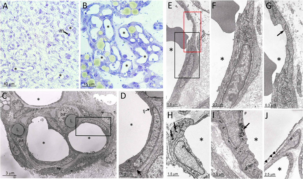
Figure 1. Murine embryonic and postnatal eWAT morphology.
A. eWAT depot at E18 composed of poorly differentiated mesenchymal cells (m = mitosis; some capillaries indicated with asterisk). B. eWAT at P7, where adipocytes appear yellowish in areas with abundant large capillaries (asterisks). C,D. EM micrograph of a vasculo-adipocytic islet showing EC (e; elongated cells), tight junctions (tj), pericytes (p; poorly differentiated cells with glycogen granules and surrounded by a distinct basal membrane), preadipocytes (pa; cells with small lipid droplets, (L)), and glycogen granules in pericyte (arrow). E. EC and pericytes of a capillary wall (asterisk indicates the capillary lumen). F. Enlargement of the black squared area in E revealing EC in endothelial-pericytic position. G. Enlargement of the red squared area in E highlighting typical oblique tight junction (arrow) joining cell in endothelial-pericytic position to adjacent EC. H. Example of cell in endothelial-pericytic position containing abundant glycogen (arrows). I. Example of a "pure" EC containing abundant glycogen (arrow). J. Example of a cell partially associated with the capillary wall (arrowheads) and partially abutting into the interstitial space.

