Figure 2.
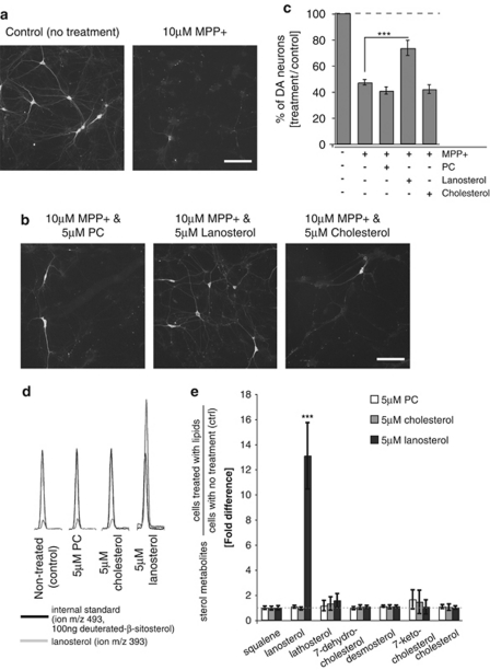
Lanosterol rescues dopaminergic neurons in MPP+-treated ventral midbrain cultures. Fluorescence images of primary ventral midbrain cultures (DIV7) stained with anti-TH, a marker for dopaminergic neurons. (a) Control cells (no treatment) and cells treated with 10 μM MPP+ (b) co-treated cells for 24 h with 10 μM MPP+ and 5 μM of phosphatidylcholine (PC, left panel), lanosterol (middle panel) or cholesterol (right panel). Scale bars in (a) and (b) represent 200 μm. (c) Plot of fold changes of each treatment relative to control. y axis shows the average percentage of TH+ neurons from each treatment condition divided by the average percentage of TH+ neurons in control. Error bars represent S.E.M. from four to five independent experiments. ***P<0.001 between lanosterol/MPP+ and MPP+. (d) GC–MS profiles of lanosterol (m/z 393, light grey) and the internal standard (deuterated β-sitosterol, m/z 493, black) in ventral midbrain cultures incubated for 24 h with various lipid treatments. (e) Cellular levels of sterol intermediates following various treatments normalized to control (no treatment). Fold changes plotted in y axis represent the levels of each metabolite in each treatment condition normalized to control. Error bars represent S.E.M. from three independent experiments. ***P<0.001 between lanosterol treatment and control

