
Figure 1.
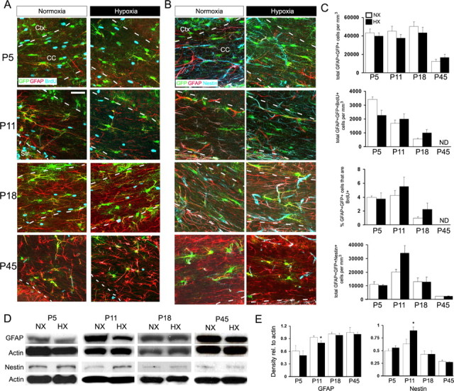
Hypoxia does not affect white matter astrocyte cell number but decreases GFAP expression and increases Nestin expression. A–C, GFAP-GFP mice were subjected to hypoxia (10.5 ± 0.5% O2) from P3 to P11 followed by an injection of BrdU 2 h before killing. Brain slices were stained with anti-GFAP (red) and anti-BrdU (blue) (A) or anti-GFAP (red) and anti-Nestin (blue) (B) antibodies. C, Quantification of percentage or number of GFAP+GFP+ cells immunostained with anti-BrdU or anti-Nestin. Scale bar, 50 μm. Cell counts are shown ± SEM. D, E, Western blot analysis of GFAP and Nestin expression in the white matter after hypoxia. Protein levels are expressed relative to actin loading control. Densitometric analysis is shown ± SD (n = 4–6 animals; *p < 0.05, unpaired Student's t test). Ctx, Cortex; CC, corpus callosum; NX, normoxia; HX, hypoxia.