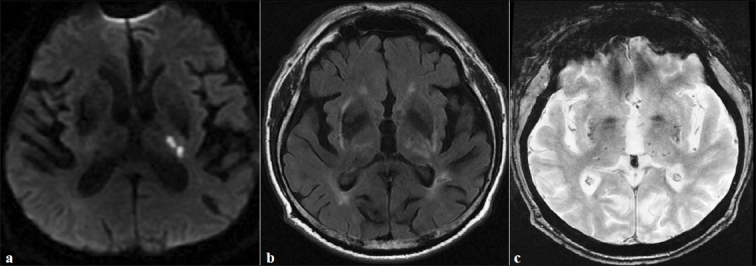
Figure 1.

(a) Diffusion-weighted MRI showing limited infarct in the left posterior arm of the internal capsule. (b) Axial FLAIR imaging showing white matter lesions and lacunar infarcts. (c) Axial gradient-echo MRI sequence showing multiple microbleeds (small foci of hypointensity) located in the basal ganglia
