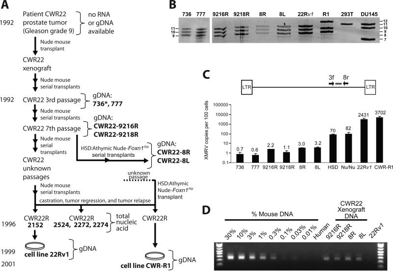
Fig. 1.
Characterization of CWR22 xenografts and XMRV-related sequences. (A) Genesis of 22Rv1 and CWR-R1 cell lines. Bold letters indicate samples from which genomic DNA (gDNA) or total nucleic acid was available for analysis. XMRV-positive samples are boxed. *, unknown early passage. (B) Short tandem repeat (STR) analysis. Representative D7S280 allele pattern of xenografts, 22Rv1 and CWR-R1 cell lines, along with analysis of six additional loci (Fig. S1). An allelic ladder is shown on left and right of gel. (C) Quantitative real-time PCR to detect XMRV env sequences. Calculated copies/100 cells are indicated above each bar. (D) IAP assay to quantitate the amount of mouse DNA present in the xenograft gDNAs.

