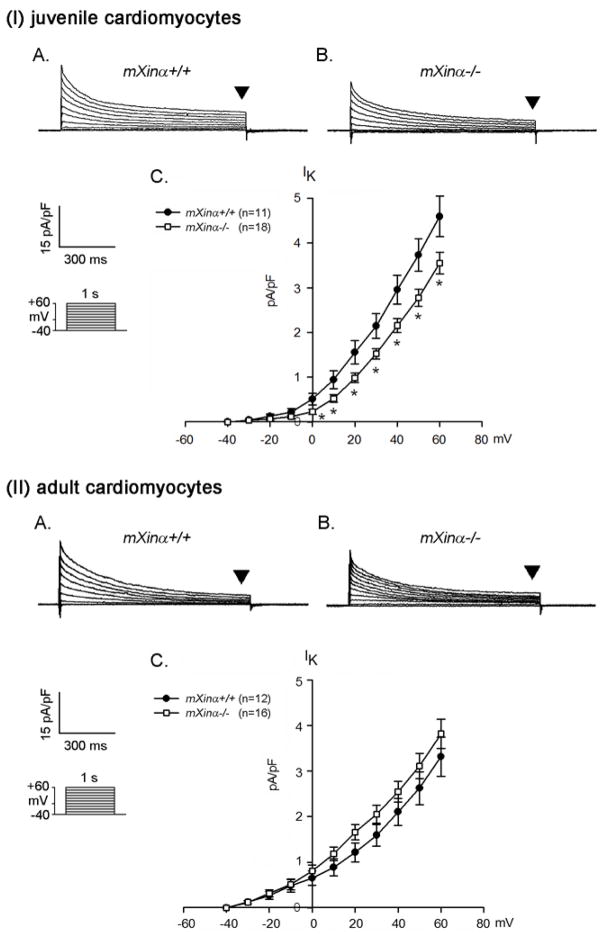
Figure 4.
The delayed rectifier outward K+ current densities of juvenile but not adult mXinα-null ventricular myocytes are significantly depressed as compared to those of wild-type counterparts. Membrane currents were elicited on depolarization from a holding potential of −40 mV to a test potential from −40 to +60 mV. Examples of delayed rectifier K+ currents (indicated by downward solid triangles, IK) recorded in the (I) juvenile and (II) adult mXinα+/+ (Panel A) and mXinα−/− (Panel B) ventricular myocytes. Inset, various clamp protocols. Panel C summarizes current density-voltage (mean±SEM) relationships of IK. n, Number of ventricular myocytes tested. * p<0.05, significant difference between juvenile wild-type and mXinα-null ventricular myocytes.

