Figure 5.
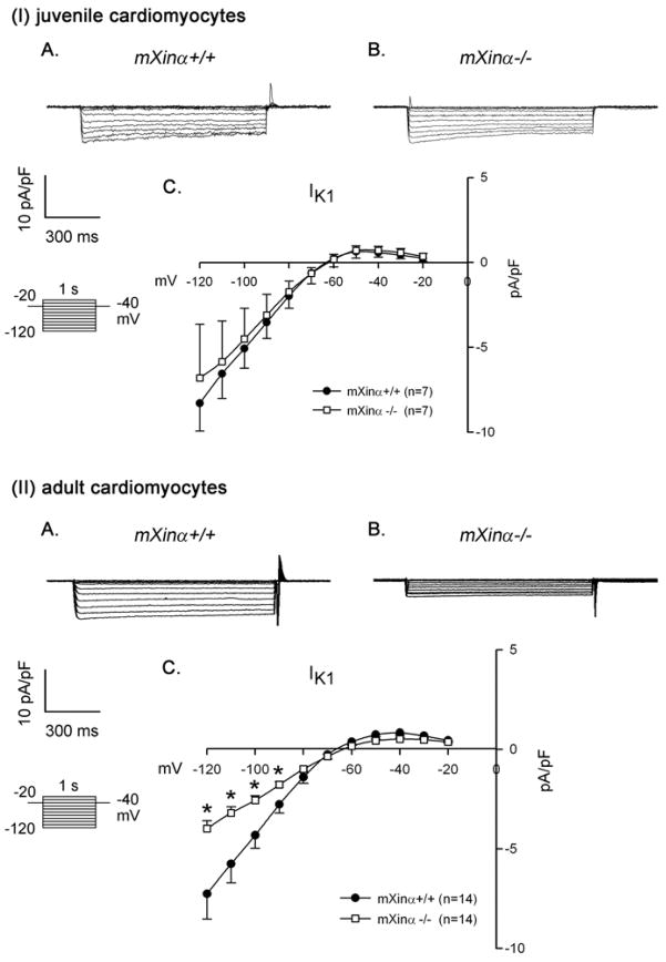
Ba2+-sensitive inward rectifier K+ current (IK1) in juvenile and adult ventricular myocytes. Top panel (I) illustrates examples of a series of IK1 currents elicited on hyperpolarization in a wild-type (mXinα+/+) (A) and a mXinα-null myocyte (B). The clamp protocol is shown below the calibration bar. The differences in current densities before and after Ba2+ (mean ± SEM) were then plotted against the test potentials to obtain current-voltage relationship curves. No change in the IK1 was detected in juvenile mXinα-null ventricular myocytes (I) as compared to juvenile wild-type myocytes (C). In contrast, the IK1 currents of adult mXinα-null ventricular myocytes (II) were significantly depressed at test potentials more negative than −90 mV as compared to the wild-type counterparts (II). * p<0.05, significant difference between wild-type and mXinα-null ventricular myocytes.

