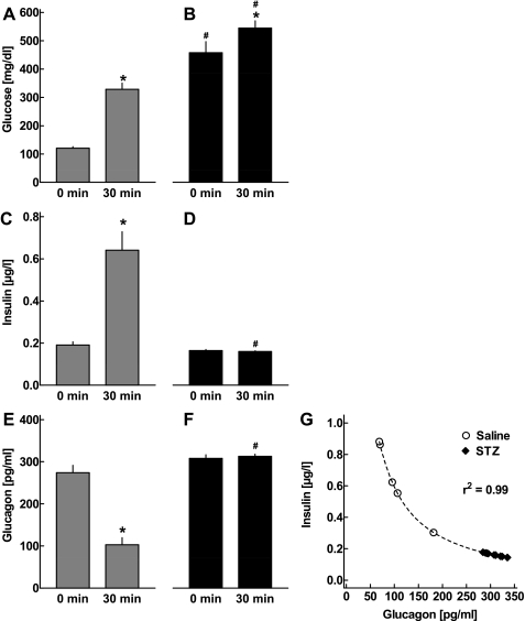
Fig. 2.
Plasma concentrations of glucose (A and B), insulin (C and D), and glucagon (E and F) in CD-1 mice 28 days after the first injection of saline (gray bars: A, C, E; n = 6) or STZ (filled bars: B, D, F; n = 13). Respective concentrations were determined in the basal state and 30 min following ip glucose injection. Glucose values exceeding the upper detection limit (600 mg/dl) were considered equal to 600 mg/dl. Data are presented as means ± SE. *P < 0.001 vs. basal levels; #P < 0.001 vs. respective concentrations in saline-treated mice. G: correlation between insulin and glucagon levels 30 min after ip glucose administration in CD-1 mice 28 days after first injection of either saline or STZ. Dashed line depicts the regression curve derived from nonlinear regression analysis (r2 = 0.99).

