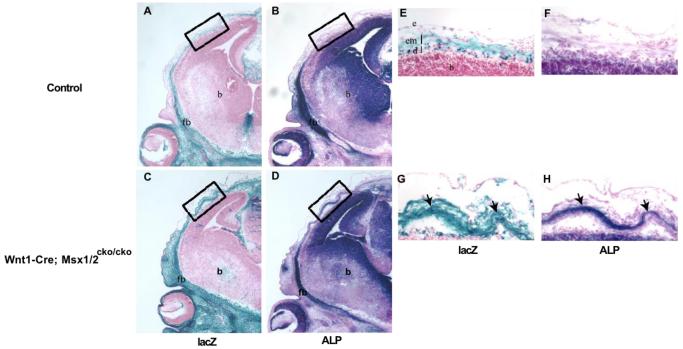
Figure 3. Neural crest origin of heterotopic bone.
To visualize neural crest-derived cells, we produced mice carrying the R26R marker allele along with Wnt1-Cre; Msx1cko/cko; Msx2cko/cko. Embryos were taken at E13.5, and heads were sectioned in the coronal plane. Adjacent sections were stained either for lacZ (A, C, E, G) or ALP (B, D, F, H). Boxed areas in A-D correspond to areas of heterotopic ALP activity (see Figure 2). Arrows in G and H point to heterotopic prospective bone. Images shown are representative of three control and three mutant embryos examined. Note that the ALP-positive cells are in a lacZ positive cell layer and are therefore derived from neural crest. fb, frontal bone; b, brain; em, early-migrating neural crest; d, dura; e, epidermis.

