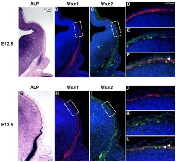
Figure 4. Expression of Msx1/2 in early migrating cranial mesenchyme.
Coronal sections of embryos at E12.5 and E13.5 were stained for ALP activity (A, G) and incubated with Msx1 (B, D, H, J) and Msx2 (C, E, I, K) probes simultaneously. Hybridization signals were visualized by immunofluorescence. Msx1 is in red, Msx2 in green. D, E, J and K show boxed areas in B, C, H and I. F and L are merges of images in D, E, J and K respectively. Note partial overlap of Msx1 and Msx2 signals in the neural crest-derived mesenchyme layer at E12.5 (yellow color, arrowhead, F). At E13.5, Msx1 is expressed in the meninges, internal to Msx2 (L, arrowheads). Msx2 is expressed in the mesenchymal layer (K).

