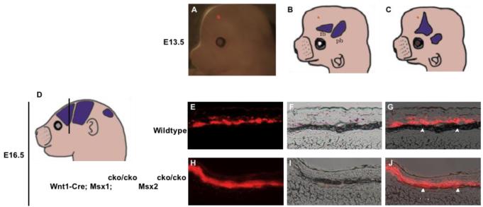
Figure 5. Inactivation of Msx1/2 in neural crest causes a change in the fate of early migrating cranial mesenchyme.
We injected DiI into heads of E13.5 control and Msx1/2 cko/cko; Wnt1-Cre and control embryos and assessed the distribution of dye after exo utero development until E16.5. Dye was placed near the apex, in the area in which the frontal bone will develop in control embryos and heterotopic bone will develop in Msx1/2cko/cko; Wnt1-Cre mutants. The placement of dye is shown in a representative embryo in A, and schematically in B and C. Embryos were allowed to develop to E16.5, and were then sectioned in the coronal plane (D, see also Fig 2) and photographed (E-J). E and H are epifluourescence images, F, I, brightfield images, G, J, merged images. In control embryos, dye was distributed in a layer of cells flanking the prospective bone. Few if any labeled cells were found in the prospective bone (arrowheads). In mutant embryos, dye was located largely in the developing bone. We obtained substantially similar results in several repetitions of this experiment (Table 1).

