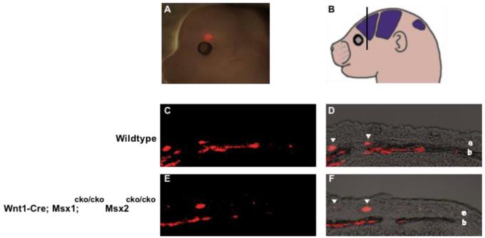
Figure 6. Osteogenic precursor cells can migrate apically from the supraorbital ridge in Msx1/2 cko/cko; Wnt1-Cre mutant embryos.
To assess the apical migration of osteogenic precursor cells, we injected DiI in the supraorbital ridge of control and mutant embryos at E13.5 as shown in A. Embryos were allowed to develop exo utero until E16.5, and were then sectioned in the indicated plane (B) and photographed. Note labeled precursor cells in the ectocranial layer (e, arrowheads) as previously described (Ting et al., 2009; Yoshida et al., 2008). These cells add to the leading edge of the growing bone (b) in both control and mutant embryos, suggesting that this morphogenetic mechanism is functional in Msx1/2cko/cko; Wnt1-Cre mutants. We obtained substantially similar results in several repetitions of this experiment (Table 1).

