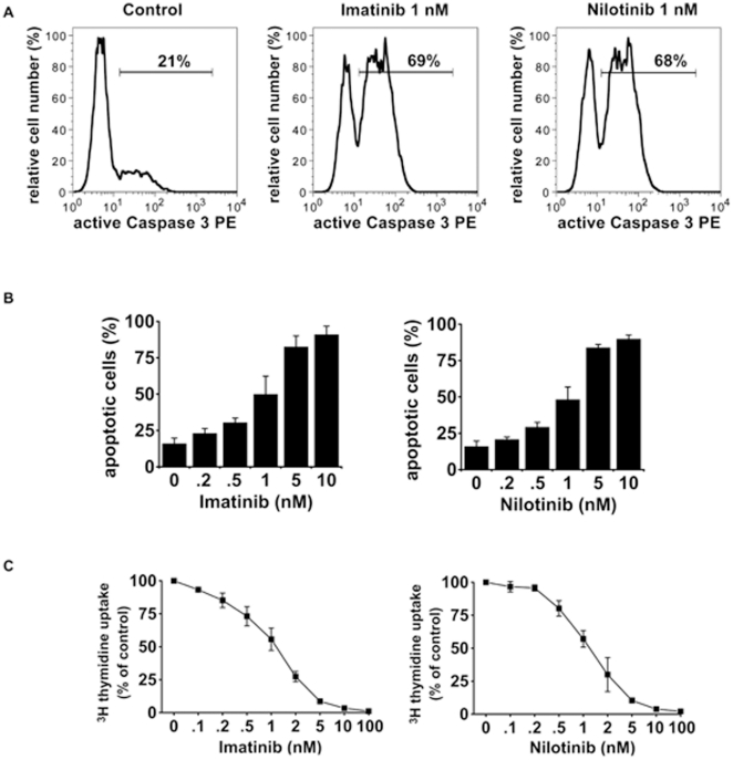
Figure 1. Induction of apoptosis and inhibition of proliferation of EOL-1 cells by imatinib and nilotinib in vitro.
(A) EOL-1 cells were cultured in medium supplemented with 1 nM imatinib (middle panel) or 1 nM nilotinib (right panel) for 48 hours. Fractions of active caspase 3 positive cells were quantified by flow cytometry. One typical experiment from three independent experiments is shown. (B) EOL-1 cells were cultured in the absence (0) or presence of various concentrations of imatinib (left panel) or nilotinib (right panel). After incubation, cells were examined for the percentage of apoptotic cells by light microscopy. Results represent the mean±S.D. from three independent experiments. (C) Dose-dependent effects of imatinib (left panel) and nilotinib (right panel) on proliferation of EOL-1 cells. Cells were kept in control medium (0) or various concentrations of imatinib or nilotinib for 48 hours. Thereafter, 3H-thymidine uptake was measured. Results show the percent 3H-thymidine uptake in drug-exposed cells relative to control (100%) and represent the mean±S.D. from 3 independent experiments.

