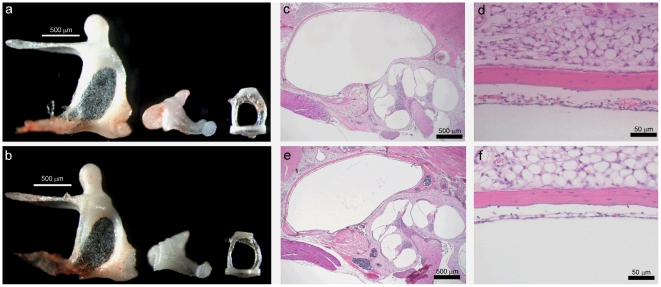
Figure 4. Middle Ears of Control and Tc1 mice.
A and B, illustrate the malleus (left), incus (middle) and stapes (right), from a wildtype mouse and a Tc1 mouse, respectively, showing no differences. C–F illustrate sections through the mucosal lining of the middle ear (H&E stained) for a wildtype (C, E) and Tc1 (D, F) mouse, showing no sign of any middle ear inflammation.

