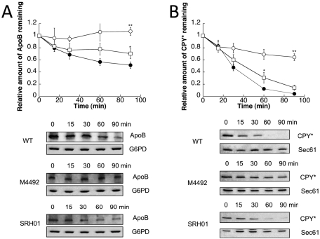
FIGURE 2:
The ERAD of ApoB29 and CPY* is slowed by the loss of PDI1. Cycloheximide chase reactions were performed as described in Materials and Methods in wild-type (●), M4492 (pdi1Δmpd1Δmpd2Δeug1Δeps1Δ [MPD1]) (○), or SRH01 (pdi1Δmpd1Δmpd2Δeug1Δeps1Δ [PDI1]) (□) yeast strains expressing ApoB29 (A) or CPY* (B). Chase reactions were performed at 30°C, and lysates were immunoblotted with anti-HA antibody. Anti-Sec61 antiserum was used as a loading control for chase reactions monitoring CPY* turnover, and anti-G6PD antiserum was used as a loading control for chase reactions measuring ApoB29 degradation. Top, quantitative data. Bottom, representative blots. Data represent the means of four to six experiments, ± SEM. The lack of visible error bars indicates that the SEM is less than the size of the symbol. *p 0.05, **p < 0.01.

