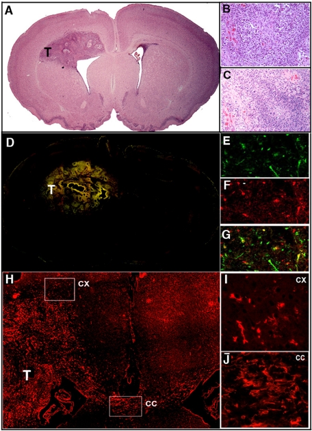
FIGURE 5:
Coinjection of EGFR-mCherry– and PDGF-IRES-GFP–encoding retroviruses into the 3-d-old rat white matter induces the formation of GBM. (A) At 10-d postinjection, a tumor mass (T) is apparent (hematoxylin and eosin staining; 4× objective). With a 20× objective, the tumor is seen to demonstrate the hallmarks of GBM, including vascular proliferation (B) and pseudopallisading necrosis (C). (D) Montage of fluorescence micrographs (4× objective) of red and green cells show that the tumor mass is composed of both green PDGF-IRES-GFP–expressing cells (20× objective in E), red EGFR-mCherry-expressing cells (20× objective in F) and yellow double-infected cells (20× objective in G). (H) Low-power (4× objective) micrograph of mCherry fluorescence demonstrates EGFR(+) cells not only within the tumor mass (T), but also invading the cortex (CX) and corpus callosum (CC). This is also illustrated with a 10× objective for the cortex (I) and corpus callosum (J).

