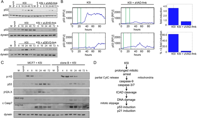
FIGURE 6:
p53 induction after mitotic DNA damage is caspase- and ICAD/CAD-dependent. (A) Immunoblotting of mock, untreated (M) and G2-synchronized (synch) cells, and cells released into K5I showed strong, persistent induction of p53 after release that was significantly blocked by caspase-9 or -7 inhibition. Actin and dynein are loading controls. (B) Live MCF7 p53-Venus cells. First vertical green line in traces is entry into mitotic arrest, second is mitotic slippage. In K5I, p53 is often induced to sustained, high levels. Cells cotreated K5I and zVAD-fmk showed only transient induction of p53 to low levels. RFU, relative fluorescence units. Caspase inhibition resulted in decreased fold induction (average of all cells ± SE) in individual cells and in fewer cells with >2 fold induction. Fold induction is the ratio of the peak intensity to the lowest intensity in each cell. K5I, n = 32; K5I + zVAD-fmk, n = 49. (C) Mock, untreated (M), MCF7, and noncleavable ICAD clone B were G2-synchronized (synch) and released into K5I. When ICAD cleavage is blocked, both p53 induction and DNA damage (γH2A.X) are decreased. Importantly, caspase-7 remains activated after K5I treatment. Dynein is loading control. All blots in each panel are from the same gels and/or samples. (D) Summary of pathway causing DNA damage during mitotic arrest that results in postslippage p53 induction.

