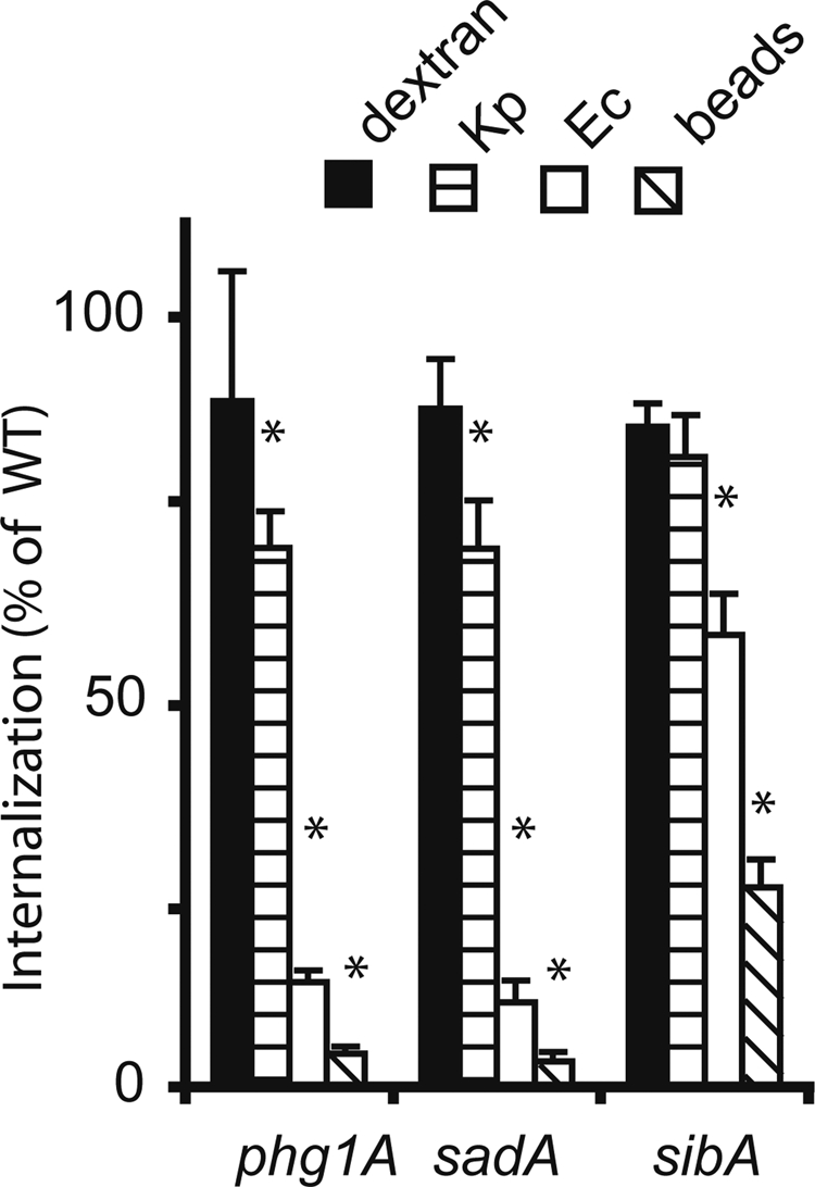
FIGURE 1:

Phagocytosis defects in phg1A, sadA, and sibA knockout cells. Wild-type or mutant Dictyostelium cells were incubated for 20 min in HL5 medium containing either fluorescent phagocytic particles (latex beads [beads], K. pneumoniae [Kp], or E. coli [Ec] bacteria) or a fluorescent fluid-phase marker (dextran). The internalized fluorescence was analyzed by flow cytometry and expressed as a percentage of internalization in wild-type cells. Qualitatively, phg1A, sadA, and sibA mutants presented similar phenotypes compared with wild-type cells: virtually normal uptake of fluid phase and of K. pneumoniae, diminished phagocytosis of E. coli, and minimal phagocytosis of latex beads. The average and SEM of at least four experiments are indicated. *, significantly different from wild-type (p < 0.05 with Student's t test).
