
FIGURE 5:
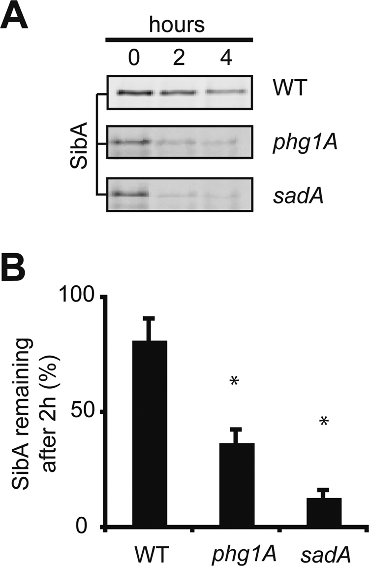
The SibA protein is less stable in phg1A and sadA mutant cells than in wild-type cells. (A) Wild-type and mutant cells were incubated in the presence of cycloheximide for 0, 2, or 4 h and then lysed; the remaining SibA was detected by Western blotting after gel electrophoresis. To obtain equivalent signals at time zero, 106 cells were loaded per lane for phg1A and sadA mutant cells and 105 for wild-type cells. (B) The signals were quantified to determine the percentage of SibA remaining after 2 h of chase. The average and SEM of six independent experiments are indicated. *, significantly different from wild-type (p < 0.05 with Student's t test).