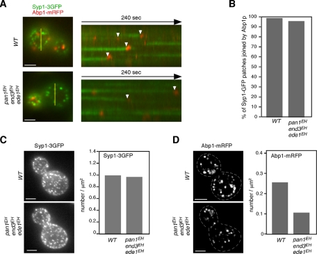
FIGURE 8:
All Syp1p patches are eventually internalized in triple mutants. (A) Epifluorescence images of a single cell coexpressing Syp1-3GFP and Abp1-mRFP. Yellow bars mark where the kymograph was generated. Right, kymograph representations of Syp1-3GFP and Abp1-mRFP from 3-min movies. Arrowheads indicate examples of Syp1p patches that are joined by Abp1-mRFP. Scale bars, 2 μm. (B) Percentage of the Syp1-3GFP patches that were eventually joined by Abp1-mRFP. (C, D) Maximum-intensity projections of Z stacks of wild-type and triple-mutant cells labeled with Syp1-3GFP (C) or Abp1-mRFP (D). The Z series was acquired through the entire cell at 0.2-μm intervals. Dotted lines represent outline of cells. Quantification of Syp1-3GFP or actin patches/μm2 ± SD in wild-type and triple mutant cells (n = 50 cells for each strain). Scale bars, 2 μm.

