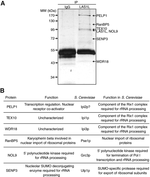
FIGURE 1:
Isolation of LAS1L-associated proteins. (A) LAS1L was affinity-purified from HEK 293T cell lysate using an anti-LAS1L antibody coupled to protein G Sepharose beads. Normal rabbit IgG was used as negative control. LAS1L-associated complexes were separated on SDS–PAGE and silver-stained and bands present only in the LAS1L IP lane were cut out, digested with trypsin, and analyzed by mass spectrometry. Identified proteins are marked with arrows. (B) The proteins identified in (A) and their corresponding S. cerevisiae putative homologues (Rout et al., 1997; Jakel and Gorlich, 1998; Bassler et al., 2001; Vadlamudi et al., 2001, 2004; Peng et al., 2003; Galani et al., 2004; Krogan et al., 2004; Gong and Yeh, 2006; Panse et al., 2006; Nair et al., 2007; Haindl et al., 2008; Yun et al., 2008; Braglia et al., 2010; Chou et al., 2010; Heindl and Martinez, 2010; Finkbeiner et al., 2011).

