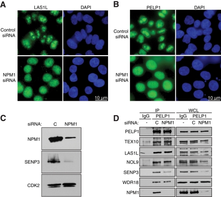
FIGURE 10:
NPM1 is required for LAS1L and PELP1 nucleolar localization. (A) HCT116 cells were transfected with control or NPM1 siRNA for 48 h. Subcellular localization of LAS1L was determined by immunofluorescence analysis using a LAS1L antibody (green). DNA was visualized by staining with Hoechst 33342 (blue). Scale is representative of all panels. (B) HCT116 cells were transfected with control or NPM1 siRNA for 48 h. Subcellular localization of PELP1 was determined by immunofluorescence analysis using a PELP1 antibody (green). DNA was visualized by staining with Hoechst 33342 (blue). Scale is representative of all panels. (C) Knockdowns of NPM1 for (A) and (B) were confirmed by Western blotting using specific antibodies (indicated on the left). An anti-CDK2 antibody was used as loading control. (D) Cells were transfected with a control (“C”) or NPM1 siRNA for 72 h. Lysates were then immunoprecipitated with rabbit IgG (as negative control) or PELP1 antibody. Coprecipitating proteins were separated on SDS–PAGE and analyzed by Western blotting using specific antibodies (indicated on the left). WCL, whole-cell lysate.

