FIGURE 2:
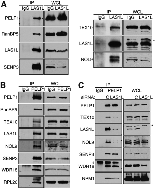
LAS1L associates with PELP1, TEX10, WDR18, RanBP5, NOL9, and SENP3. (A) LAS1L was immunoprecipitated (IP) with an anti-LAS1L–specific antibody from HEK 293T cell lysates. Associated proteins were separated on SDS–PAGE and analyzed by Western blotting with specific antibodies (indicated on the left). Normal rabbit IgG was used as negative control. WCL, whole-cell lysate; *, presence of an unspecific band. (B) PELP1 was immunoprecipitated (IP) with an anti-PELP1 antibody from HEK 293T cell lysates. Associated proteins were separated on SDS–PAGE and analyzed by Western blotting with specific antibodies (indicated on the left). Normal rabbit IgG was used as negative control. WCL, whole-cell lysate. (C) HEK 293T cells were transfected with nontargeting control (represented by the letter “C”) or LAS1L siRNA for 48 h. Cells were lysed and immunoprecipitated with an anti-PELP1 antibody. Proteins associating with PELP1 in the absence of LAS1L were separated on SDS–PAGE and analyzed by Western blotting with specific antibodies (indicated on the left). Normal rabbit IgG was used as negative control. WCL, whole-cell lysate; *, presence of an unspecific band; #, an IgG band.

