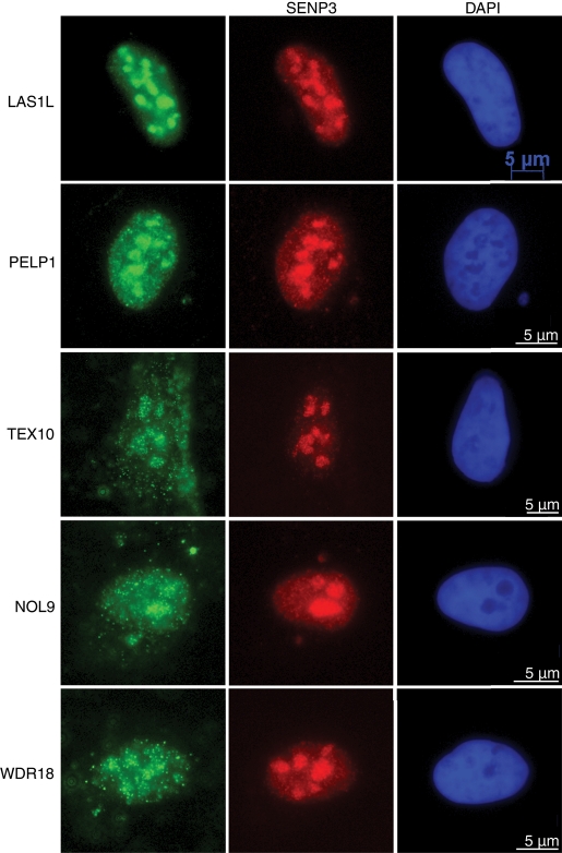
FIGURE 4:
LAS1L-associated proteins localize to the nucleolus. Immunofluorescence analysis of U2OS cells with complex protein-specific antibodies. Cells were preextracted with 0.1% Triton, fixed, and immunostained with anti-LAS1L, PELP1, TEX10, NOL9, and WDR18 antibodies (green). Colocalization with SENP3 was confirmed using an anti-SENP3 antibody (red). DNA was visualized by staining with Hoechst 33342 (blue). Scale is representative of all three panels.

