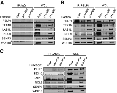
FIGURE 6:
LAS1L and NOL9 interact with the mammalian Rix1 complex on pre-60S ribosomal particles. Nuclear extracts from HCT116 cells were fractionated by centrifugation on a 10–30% sucrose gradient. Fractions were collected, and the optical density was measured at 260 nm (A260). Based on the A260 profile, fractions corresponding to free nuclear proteins (1, 2, and 3), pre-40S ribosomal particles (8, 9, and 10), and pre-60S ribosomal particles (12, 13, and 14) were then combined and immunoprecipitated (IP) with rabbit IgG (A) as a negative control or with PELP1 (B) or LAS1L (C) antibodies. Associated proteins were separated on SDS–PAGE and analyzed by Western blotting with specific antibodies (indicated on the left). WCL, whole-cell lysate; *, presence of an unspecific band; #, an IgG band.

