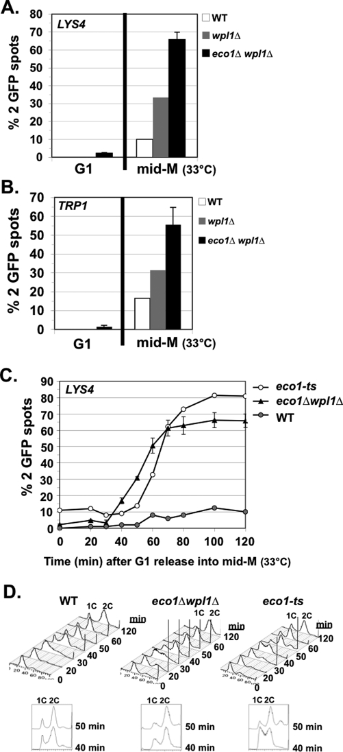
FIGURE 3:
eco1Δ wpl1Δ cells are defective in cohesion at CEN-proximal and distal loci. The percentage of cells with two GFP signals is plotted. Cells released from G1 phase were arrested in mid M phase at 33°C. (A) Cohesion loss at a CEN-distal locus (LYS4) in mid M phase. Haploids VG3349-1B (WT), VG3360-3D (wpl1Δ), VG3503-1B (eco1Δ wpl1Δ), and VG3503-4A (eco1Δ wpl1Δ). (B) Cohesion loss at CEN-proximal locus (TRP1) in mid M phase. Haploids VG3460-2A (WT), VG3513-1B (wpl1Δ), VG3502-2A (eco1Δ wpl1Δ), and VG3503-4C (eco1Δ wpl1Δ). (C) Kinetics of cohesion loss at a CEN-distal locus (LYS4). Haploids VG3349-1B (WT), VG3506-5D (eco1-ts), VG3503-1B (eco1Δwpl1Δ), and VG3503-4A (eco1Δ wpl1Δ) assayed for cohesion loss after release from G1 phase. (D) FACS. Data were derived from two independent experiments, and error bars are SD. For cohesion assays,100–400 cells were scored for each data point in each experiment.

