FIGURE 6:
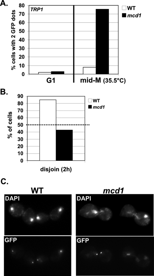
Random segregation of sister chromatids in mcd1-1 cells released from nocodazole arrest. WT (VG3460-2A) and mcd1-1 mutant (VG3456-2C) cells arrested in mid M phase 35.5°C using nocodazole and then released from arrest (23°C). Chromosome IV monitored at a CEN4-proximal locus (TRP1). (A) Cohesion at mid-M-phase arrest. Percentage of cells with two GFP signals plotted for G1 and mid-M-phase cells. (B, C) Large-budded cells assayed 2 h after release from nocodazole. (B) Disjunction of chromosome IV sisters. Dotted line marks 50% disjunction expected for random segregation. (C) Micrographs showing chromosomal DNA (DAPI) and CEN-proximal locus of chromosome IV sisters (GFP). For cohesion and nondisjunction assays, 200–400 cells were scored for each data point.

