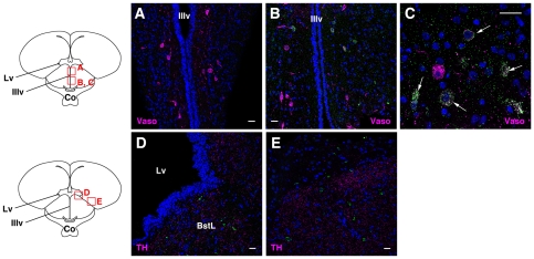
Figure 5. Distribution of cOpn5L2 immunoreactive neurons in the forebrain.
A–C, Coronal sections through the anterior hypothalamus at P10. A, The dorsal part of the paraventricular nucleus, in which many vasotocin-positive cells are seen, but no cOpn5L2 IR cells are observed. B, Paraventricular nucleus, ventral to the region shown in (A), in which cOpn5L2 IR cells are seen. C, High magnification of paraventricular nucleus. A different section from that shown in (B). Some vasotocin IR cells are also positive for cOpn5L2 (arrows). D, E, Coronal sections through the anterior hypothalamus. Images are focused on the telencephalon. D, cOpn5L2 IR cells are found in the bed nucleus of the stria terminalis, lateral part (BstL), in which TH IR fibers are prominent. E, cOpn5L2 IR cells are also scattered in the lateral region of the telencephalon. Co, optic chiasm; IIIv, third ventricle; Lv, lateral ventricle. The scale bars represent 20 µm.

