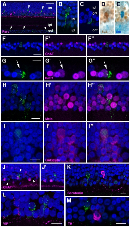
Figure 6. Immunohistochemistry of the chick retina at P10.
A, cOpn5L2 IR cells in the inner nuclear layer (inl) and ganglion cell layer (gcl) (green, arrowheads). Parvalbumin is visualized in magenta to reveal subsets of amacrine cells in the inner nuclear layer (inl) and sublamina I and V in the inner plexiform layer (ipl) [47]. B, A representative cOpn5L2 IR cell in the INL. C, A representative cOpn5L2 IR cell in the GCL. D, A cOpn5L2 IR cell in the INL after two-color ABC immunostaining. E, A cOpn5m IR cell in the INL of the same retinal section as shown in (D). F-F″, The cOpn5L2 IR cell (green in F, F″) in the GCL is not positive for ChAT (magenta in F′, F″). G-G″, The cOpn5L2 IR cell (arrow) in the GCL (G, G″) is positive for Islet1 (G′, G″). H-H″, cOpn5L2 IR cells in the INL (H, H″) are positive for Meis (H′, H″). I-I″, The cOpn5L2 IR cell in the INL (I, I″) is positive for GAD65/67 (I′, I″). J, J′, cOpn5L2 IR cells in the INL (green, arrowheads in J) are not positive for ChAT. Some cOpn5L2 IR cells are adjacent to ChAT IR cells (J), and others are separate from the ChAT IR cells (J′). K, One cOpn5L2 IR cell in the vicinity of a serotonin IR cell, while the other is located apart from it. L, cOpn5L2 IR cells are not positive for VIP. M, A cOpn5L2 IR cell adjacent to a TH IR cell. For all images, DAPI is blue. Scale bars, 50 µm (A), 10 µm (B–E, G-I″, K–M), and 20 µm (F-F″, J, J′).

