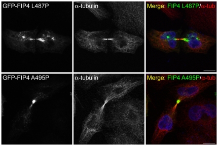
Figure 7. FIP4 localises to the midbody of dividing cells independently of TSG101.
HeLa cells were transfected with constructs encoding the indicated proteins. At 16–18 hours post-transfection, cells were processed for immunofluorescence microscopy and immunostained for α-tubulin. DAPI was used to visualise the nuclei. Images were acquired by confocal microscopy. Scale bar indicates 10 µm. Data are typical of at least three independent experiments.

