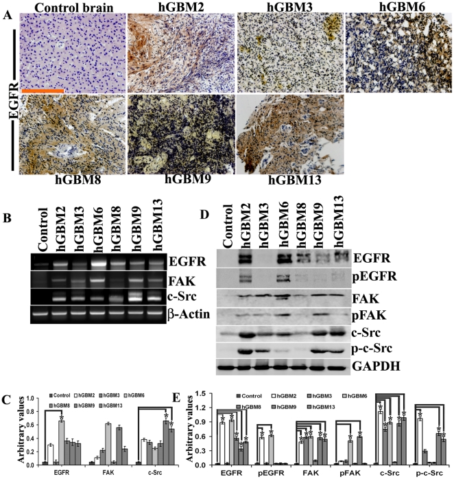
Figure 1. Expression of EGFR in GBM-patient derived specimens.
(A) Control brain and GBM-patient derived specimens were subjected to DAB immunohistochemistry using anti-rabbit EGFR antibody (bar = 200 µm). (B) mRNA extracted from the above specimens was subjected to RT-PCR using primers specific for EGFR, FAK, c-Src and β-actin (loading control). (C) Quantitative analysis of (B). (D) Tissue lysates (40 µg protein) of the above specimens were subjected to Western blotting using the following antibodies: EGFR, pEGFR, FAK, pFAK, c-Src and phospho-c-Src. Mouse anti-GAPDH (1∶1000) served as the loading control. (E) Quantitative analysis of (D). *Significant at p<0.05 compared to control brain samples.

