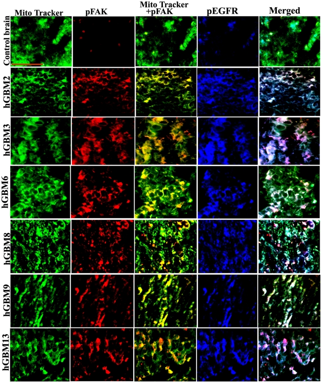
Figure 2. Colocalization of pEGFR and pFAK in clinical samples.
Control brain and GBM-patient derived specimens were labeled with pEGFR and pFAK antibodies along with Mito Tracker (green) and processed for immunofluorescence. pEGFR was conjugated with Alexa Fluor 350 (blue) and pFAK was conjugated with Alexa Fluor 594 (red) secondary antibodies (bar = 100 µm).

