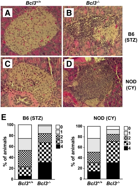
FIG. 2.
Histologic profiles of pancreas. Mice were treated as described in Fig. 1 and killed 45 days after the first streptozotocin (STZ) or cyclophosphamide (CY) injection. Pancreata were collected, fixed in 10% formalin, and embedded in paraffin. Paraffin sections (5-μm thick) were stained with hematoxylin and eosin. Pancreatic sections of B6 (A) and B6xNOD (C) Bcl3+/+ mice showed little or no insulitis, whereas pancreatic sections of B6 (B) and B6xNOD (D) Bcl3−/− mice showed severe insulitis. Magnification: ×200. E: Insulitis scores. Mice were treated as in Fig. 1, and pancreatic inflammation was graded as follows: 0, no inflammation; 1, peri-insulitis with mononuclear cell infiltration affecting 25% of the circumference; 2, peri-insulitis with mononuclear cell infiltration affecting 25% of the circumference; 3, mild-to-moderate insulitis with intraislet mononuclear cell filtration but good preservation of islet architecture; 4, severe insulitis with numerous intraislet inflammatory cells and loss of normal islet architecture. (A high-quality digital representation of this figure is available in the online issue.)

