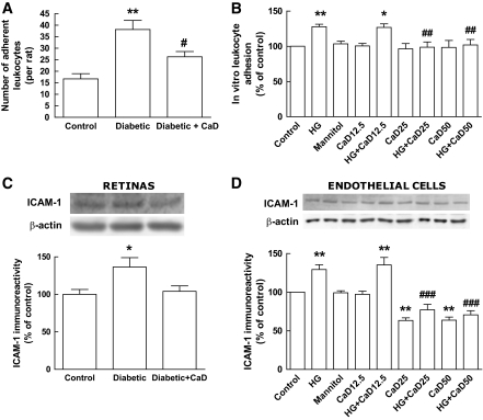
FIG. 4.
Diabetes and elevated glucose increase the number of leukocytes adhering to retinal vessels and retinal endothelial cells and the content of ICAM-1: protective effect of CaD. A: Quantification of leukocytes adhering to retinal vessels. Data are presented as number of adherent leukocytes to retinal vessels per rat (two retinas) and represent the mean ± SEM of seven animals. B: Quantification of leukocyte adhesion to retinal endothelial cells (TR-iBRB2 cell line) using a fluorometric assay. Data are presented as percentage of control and represent the mean ± SEM of 7–10 independent experiments. C: The protein levels of ICAM-1 were evaluated in whole rat retinal extracts by Western blotting. Data are presented as percentage of control and represent the mean ± SEM of seven animals. D: The protein levels of ICAM-1 were evaluated in whole extracts of rat retinal endothelial cell cultures (TR-iBRB2 cell line) by Western blotting. Data are presented as percentage of control and represent the mean ± SEM of at least four independent experiments. *P < 0.05, **P < 0.01, significantly different from control; ANOVA (one-way) followed by Dunnett post hoc test. #P < 0.05, ##P < 0.01, ###P < 0.001, significantly different from diabetic rat or high glucose condition; ANOVA (one-way) followed by Bonferroni post hoc test. HG, high glucose (30 mmol/l, for 4 days); CaD12.5, CaD25, and CaD50, calcium dobesilate 12.5, 25, and 50 μg/ml, respectively (4 days).

