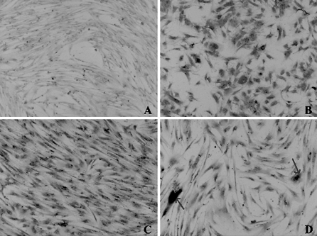
Fig. 5.
Immunocytochemistry staining for EPO protein on hMSC nucleofected with MIDGE-EPO. Cells nucleofected with MIDGE-EPO were stained for EPO protein using monoclonal antibody anti-human EPO with a Dako LSAB kit. A Staining on cells without nucleofection. B Cells were positively stained 24 h post-nucleofection and C 3 months later. D Some cells showed very strong positive staining for EPO protein (arrows). Magnification ×40

