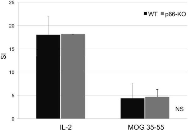
Figure 6. T cell proliferation studies showed no significant differences between p66-KO and WT mice in MOG immunoreactivity.
Using standard EAE protocol, p66-KO and WT mice were immunized with MOG 35-55 peptide. Ten days post-immunization, lymph nodes were collected for proliferation studies. Lymph node cultures treated with positive control IL-2 and MOG 35-55 peptide (25mg/mL) showed no significant differences in proliferative responses between the p66-KO and WT groups. SI = Stimulation index; NS = difference not significant (p>0.05).

