
Figure 2. Introduction to Epidermolysis Bullosa Simplex.
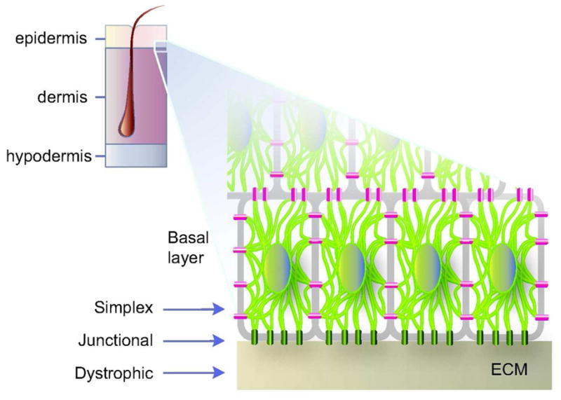
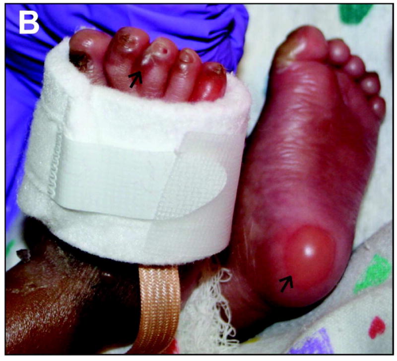
A) Schematic representation of skin tissue (left) and detailed view of the bottom portion of the epidermis (right), highlighting the cytoplasmic network of keratin IFs (light green) attached to hemidesmosome cell-ECM (green) and desmosome cell-cell (magenta) contacts in basal keratinocytes. Arrows depict the plane of tissue rupture seen in the “simplex”, “junctional”, and “dystrophic” forms of EB. B) Example of trauma-induced bullous skin lesions (arrows) in the feet of a 2-month old child diagnosed with EBS. Picture kindly provided by Dr. Bernard Cohen (Johns Hopkins School of Medicine; see http://dermatlas.med.jhmi.edu). Adapted from (Coulombe et al., 2009).