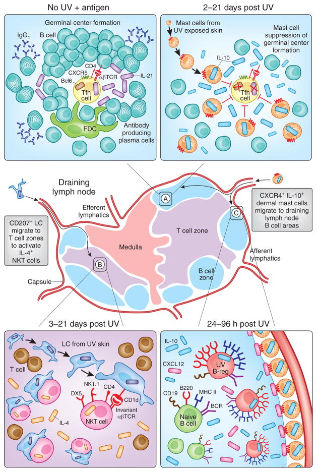
Figure 2. Photoimmunological Events Occurring in Skin Draining Lymph Nodes.
Immunosuppressive signals generated in the skin are transmitted by Langerhans Cells (LC) and mast cells to the local skin-draining lymph nodes to regulate both humoral and cell-mediated immune responses. (A: left hand panel) In response to immunization with protein antigens, B are activated by IL-21-expressing T follicular helper (Tfh) cells in germinal centers to produce IgG1 antibodies. (A: right hand panel) Mast cells that have migrated from UV-irradiated skin suppress this humoral arm of adaptive immunity by homing to B cell areas and producing IL-10 (Chacon-Salinas et al., 2011). At the same time, (B) CD207 (Langerin)+CD1d+ epidermal Langerhan’s Cells (LC) migrate from UV irradiated skin to the T cell zones where they activate IL-4-producing immunosuppressive NK-T cells (Fukunaga et al., 2010). Meanwhile, (C) UV-induced upregulation of CXCL12 (SDF1α) in B cell follicles attracts CXCR4+ dermal mast cells to the draining lymph nodes (Byrne et al., 2008). It is at this time that IL-10 producing UV-activated B regulatory cells, or “UV-B-regs” are induced (Byrne and Halliday, 2005) via a PAF and serotonin dependent mechanism (Matsumura et al., 2006).

