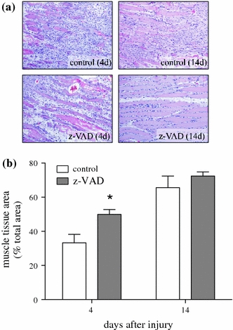
Fig. 4.

Representative light microscopic images (a) of HE staining (×100 magnification) as well as quantitative analysis of muscle tissue area (b). Animals underwent a standardized open crush injury to the left soleus muscle and treatment with DMSO as vehicle solution (control white bars, n = 6 animals per time point) or pan-caspase inhibitor z-VAD.fmk (z-VAD gray bars, n = 6 animals per time point). Analysis was performed at days 4 and 14 after injury. Data are given as means ± SEM; t-test: *P < 0.05 versus control (refer text for exact P values)
