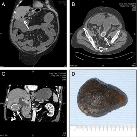
Figure 2.
CT shows a chole-colic fistula,with air seen in the biliary tree (A), dilated fluid filled proximal sigmoid, collapsed distal sigmoid and a gallstone at the transition point (B), an earlier CT showing the gallstone in the gallbladder (C) and a picture of the gallstone after surgical removal (D).

