
Figure 6.
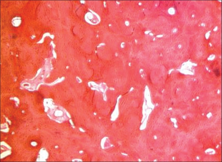
Photomicrograph shows the presence of compact lamellar bone with haversian canal, lacunae, histiocytes and reversal and resting lines suggestive of osteoma. (Hematoxylin-Eosin stain 10×).
Official websites use .gov
A
.gov website belongs to an official
government organization in the United States.
Secure .gov websites use HTTPS
A lock (
) or https:// means you've safely
connected to the .gov website. Share sensitive
information only on official, secure websites.
Photomicrograph shows the presence of compact lamellar bone with haversian canal, lacunae, histiocytes and reversal and resting lines suggestive of osteoma. (Hematoxylin-Eosin stain 10×).