Figure 10.

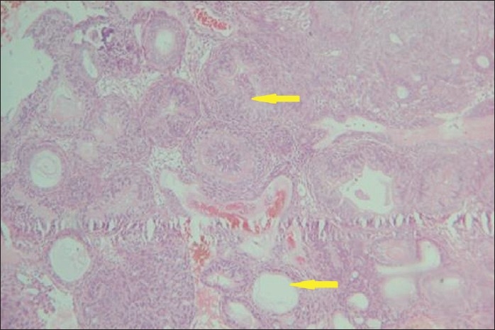
Hematoxylin and Eosin stained tissue (10×) shows odontogenic epithelial lining arranged in a rosette, ductal, and whorling pattern (arrows).

Hematoxylin and Eosin stained tissue (10×) shows odontogenic epithelial lining arranged in a rosette, ductal, and whorling pattern (arrows).