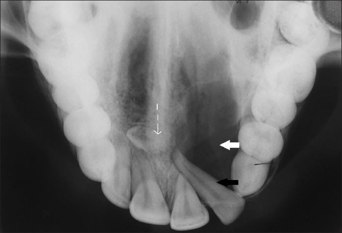
Figure 8.

Maxillary occlusal view reveals unilocular radiolucency with a sclerotic border in relation to the maxillary left central incisor to canine (teeth 21-23) region (white arrow), displaced maxillary left lateral incisor (tooth 22, black arrow), and presence of impacted supernumerary teeth in the midline (dashed arrow).
