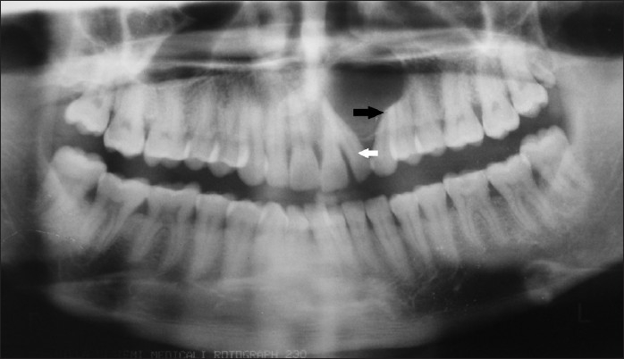
Figure 9.

Orthopantomograph (OPG) reveals unilocular radiolucency with an impacted supernumerary tooth in the midline, with displaced maxillary left lateral incisor (white arrow) and root resorption in the maxillary left canine and premolar (black arrow).
