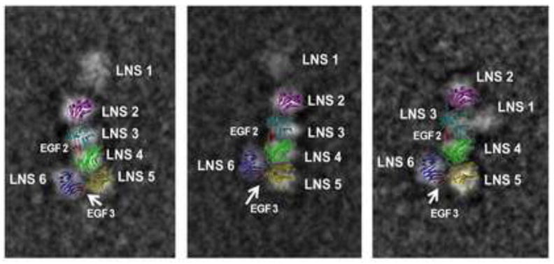
Figure 6. 2-D overlay of the α-NRXN_2-6 structure on negative stain EM images.

A 2-D image of the α-NRXN_2-6 structure overlaid onto three previously published 2-D negative stain single particle EM images of the α-NRXN_1-6 purified protein (Comoletti et al., 2010). The overall arrangement of the LNS 2-6 domains is conserved and the variable placement of the flexible LNS 1 domain relative to the crystal structure is observed.
