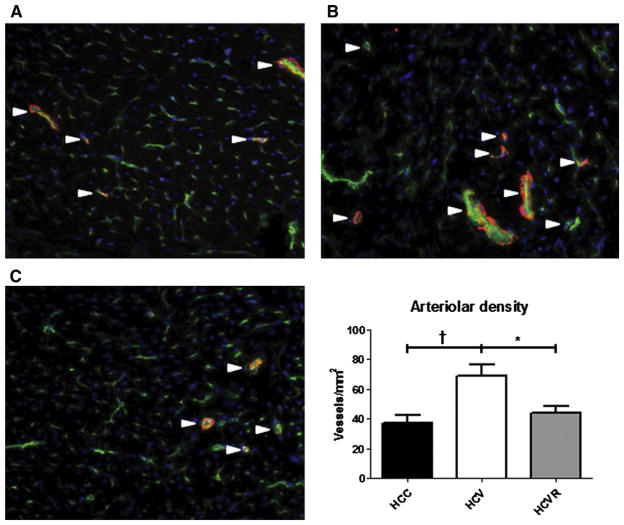
Fig 4.
Arteriolar density. Sections from the AAR were stained for endothelium-specific CD31 (green), smooth muscle actin (red), and nucleus-specific DAPI (blue). Shown are representative sections from (A) HCC, (B) HCV, and (C) HCVR animals. Arterioles were defined as structures co-staining for both CD-31 and smooth muscle actin (white arrows). Arteriolar density was significantly greater in the HCV group compared with both other groups. *P < .05; †P < .01.

