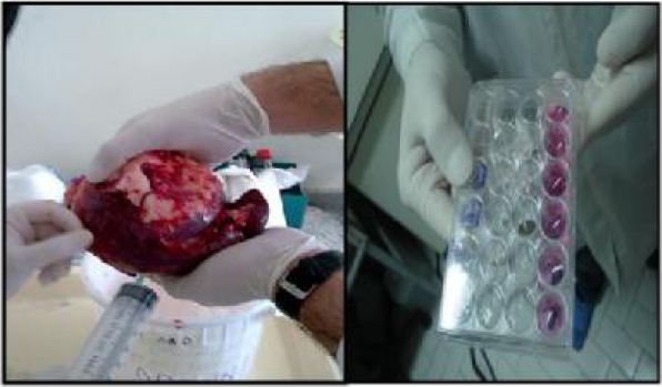
Fig.1.
Left: Aspiration of hydatid fluid from a splenic cyst. Right: 24-well cell culture plate. From the top: Fertile hydatid fluid treated lymphocytes, Infertile hydatid fluid treated lymphocytes, Cell control (all for measurement of caspase 3 activity after 6 hours), Fertile hydatid fluid treated lymphocytes, Infertile hydatid fluid treated lymphocytes, Cell control (all for assessment of Bax , and Bcl-2 expression after 12 hours)

