
Figure 6.
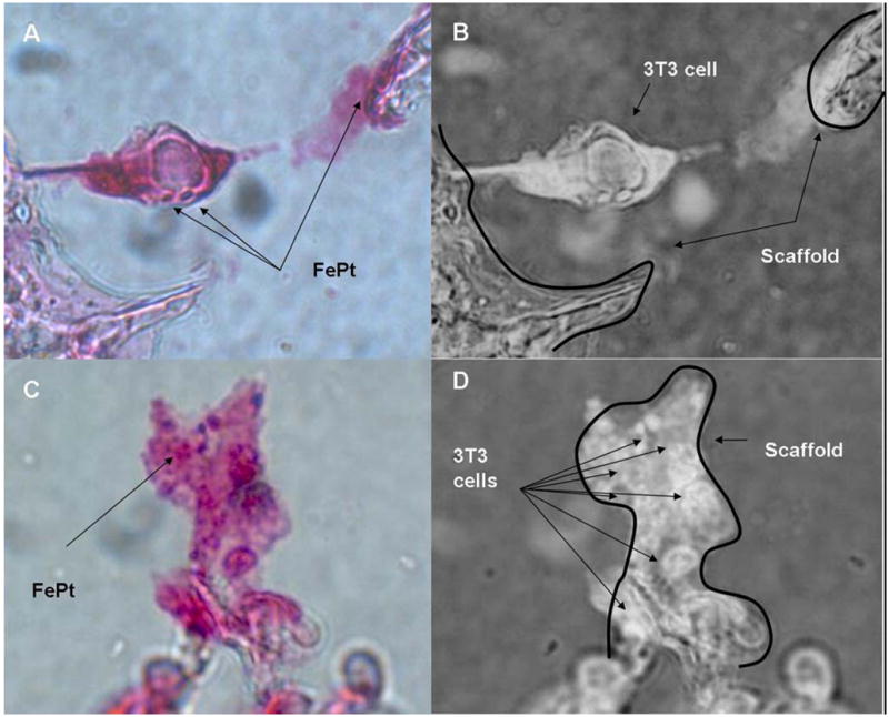
Presence of FePt particles with cells in the center section magnet applied scaffolds. (A) Prussian blue stain with counterstain of Nuclear Fast Red with positive stained FePt particles indicated as blue. (B) Inverse of (A) indicating the 3T3 cell spanning between two portions of the scaffold indicated by arrows. (C) Group of cells adhered along the scaffold with Prussian Blue stained FePt visible on the cluster of cells. (D) Inverse of image (C) showing the structure of the scaffold with arrows pointing toward the individual cells in the cluster.